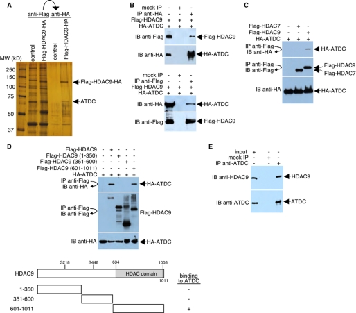
FIGURE 1.
HDAC9 interacts with ATDC. A, silver-stained SDS-PAGE of the immunopurified FLAG- and HA-tagged HDAC9-containing complexes. Control, immunopurified samples prepared in parallel from HeLa cells expressing the GFP protein. B, HeLa cells were transfected with plasmids encoding FLAG-tagged HDAC9 and HA-tagged ATDC. Whole cell lysates were immunoprecipitated and probed with FLAG-specific or HA-specific antibodies as indicated. C, HeLa cells were transfected with plasmids encoding FLAG-tagged HDAC7 or HDAC9 and HA-tagged ATDC. Whole cell lysates were immunoprecipitated and probed with FLAG-specific or HA-specific antibodies as indicated. D, top, HeLa cells were transfected with plasmids encoding full-length or deletion mutants of FLAG-tagged HDAC9 and HA-tagged ATDC. Western blot analysis was performed on the anti-FLAG immunoprecipitates with antibodies specific for HA. Levels of FLAG-tagged HDAC9 and HA-tagged ATDC were determined by Western blot analysis of cell extracts using antibodies specific for FLAG or HA, respectively. Bottom, a schematic diagram (not drawn to scale) of HDAC9 and three HDAC9 deletion mutants. Ser218 and Ser448 (S218 and S448) are serine phosphorylation sites. For simplicity, the FLAG portions of the fusion proteins are not shown. The ability of each FLAG-HDAC9 fusion protein to bind HA-ATDC is indicated (plus and minus signs). E, HeLa cell lysates were incubated with preimmune serum (mock IP) or the anti-ATDC antibody. Precipitated materials were analyzed by Western blotting using either anti-HDAC9 or anti-ATDC antibodies. IP, immunoprecipitation; IB, immunoblot.

