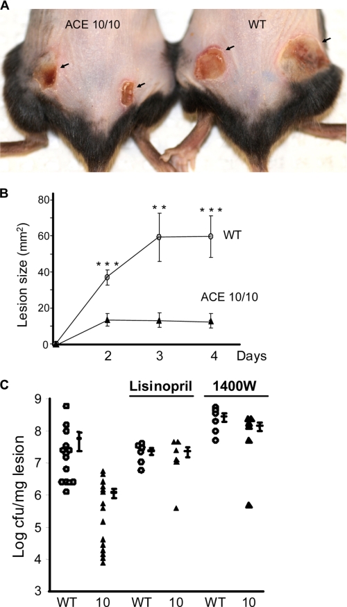
FIGURE 5.
Skin infection with MRSA. Mice were infected subcutaneously with 1 × 10⁹ MRSA, clone USA 300, in the hind flanks. A, a representative comparison of skin lesions present in ACE 10/10 and WT mice 4 days after MRSA infection. B, lesion size of WT (circles) and ACE 10/10 (10, triangles) were measured on days 2, 3, and 4 after infection (n ≥ 14 mice per group). **, p < 0.005; ***, p < 0.0005. C, four days after skin infection, WT (circles) and ACE10/10 (triangles) were sacrificed, and bacterial counts in the lesion were determined. ACE 10/10 mice averaged >50-fold less bacteria within lesions (n ≥ 14, p < 0.001). These differences were eliminated when mice were treated with either the ACE inhibitor lisinopril or the iNOS inhibitor 1400W.

