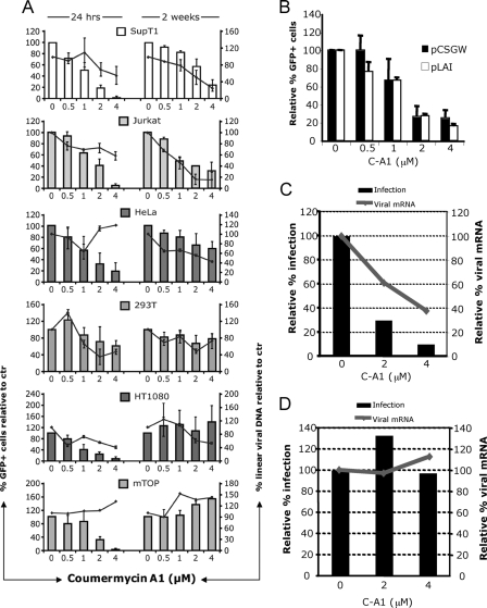
FIGURE 3.
C-A1 exerts an additional block to HIV-1 infection. A, SupT1 (human CD4+ lymphoblastic leukemia), Jurkat (human CD4+ acute lymphoblastic leukemia), HeLa (human epithelial cervix adenocarcinoma), 293T (human embryonic kidney epithelial), HT1080 (human fibrosarcoma), and mTOP (mouse fibroblasts) cells were infected with a VSV-G pseudotyped HIV-1 vector (at m.o.i. of 0.05) and analyzed by FACS (histograms, left y axis) and TaqMan qPCR (line, right y axis) 24 h and then again 2 weeks post-infection. Mean values ± S.D. are shown, n = 3. B, 293T cells transfected with HIV-1 LAIΔenv or the HIV-1 vector plasmids in the presence of the indicated doses of C-A1 and analyzed by FACS 24 h post-transfection. Mean values ± S.D. are shown, n = 3. C, HT1080 cells were infected with VSV-G pseudotyped HIV-1 LAIΔenv and analyzed 24 h later by FACS (bars, left y axis) or by RT-qPCR to measure viral mRNA levels (line, right axis). Viral RNA values were normalized to 18 S RNA copy number. D, SupT1 cells chronically infected with VSV-G pseudotyped HIV-1 LAIΔenv (at m.o.i. of 0.1) were incubated with C-A1 and analyzed 24 h later by FACS (bars, left y axis) or by RT-qPCR to measure viral mRNA levels (line, right axis). Viral RNA values were normalized to 18 S RNA copy number. Control reactions without RT gave undetectable readings. Results are representative of two independent experiments.

