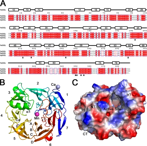
FIGURE 3.
β-Propeller fold of YeOGL. A, structural alignment of OGLs from Y. enterocolitica (YeOGL), E. chrysanthemi (EcOGL), and V. parahemeolyticus (VpOGL). The secondary structure elements of YeOGL are displayed above the aligned primary structure. Amino acids targeted for mutagenesis are indicated with black triangles below the sequences. B, schematic representation of YeOGL structure colored from the N terminus (blue) to C terminus (red). The seven β-propellers are labeled (1–7) with each strand (A–D) packing from the core toward the surface. The three bound metal ions are displayed as spheres with the catalytic manganese colored in purple and the surface-bound calciums in light blue. C, electrostatic surface potential of YeOGL. The protein is centered to display the entrance to the active site that is lined with several basic patches.

