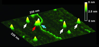
Fig. 4.
Surface plot of a Taq MutS–783Tbulge DNA complex. White arrows point to free MutS dimers on the surface. Red and blue arrows point to two dimers in a MutS tetramer bound to the DNA. The horizontal distance between the peaks of those two dimers is 9 nm. The vertical distance between the peaks of those two dimers is 0.7 nm.

