FIGURE 4.
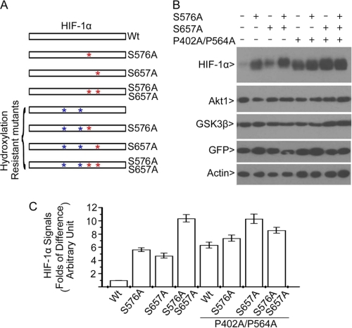
Phosphorylation of HIF-1α by Plk3 leads to its destabilization. A, schematic representation of HIF-1α and its mutants (phospho- and/or hydroxylation mutants) used for transfection analyses is shown. B, HEK293 cells were co-transfected with various expression constructs as shown in C, and a GFP expression construct for normalization of transfection efficiency for 1 day is presented. Equal amounts of cell lysates were Western blotted for HIF-1α, Akt1, GSK3β, GFP, and β-actin. C, the signals shown in B were quantified by densitometry.

