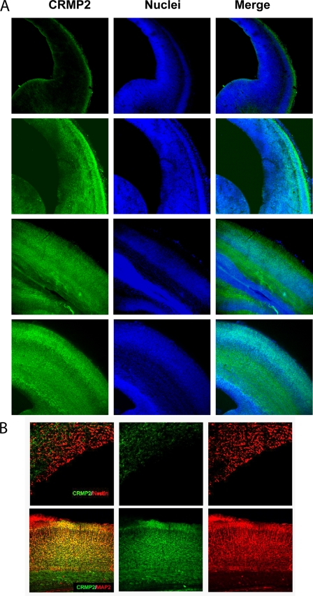
FIGURE 1.
Expression pattern of CRMP2 during cortical development. A, brain slices from E14, E16, E18, and E16 rats were subjected to immunofluorescent staining for CRMP2 and propidium iodide staining for the nuclei. B, double immunofluorescent staining of both CRMP2 and the progenitor cell marker nestin in E20 rat brain (upper panels) or of CRMP2 and mature neuron marker MAP2 in E18 mouse brain to determine the identity of CRMP2-positive cells.

