FIGURE 3.
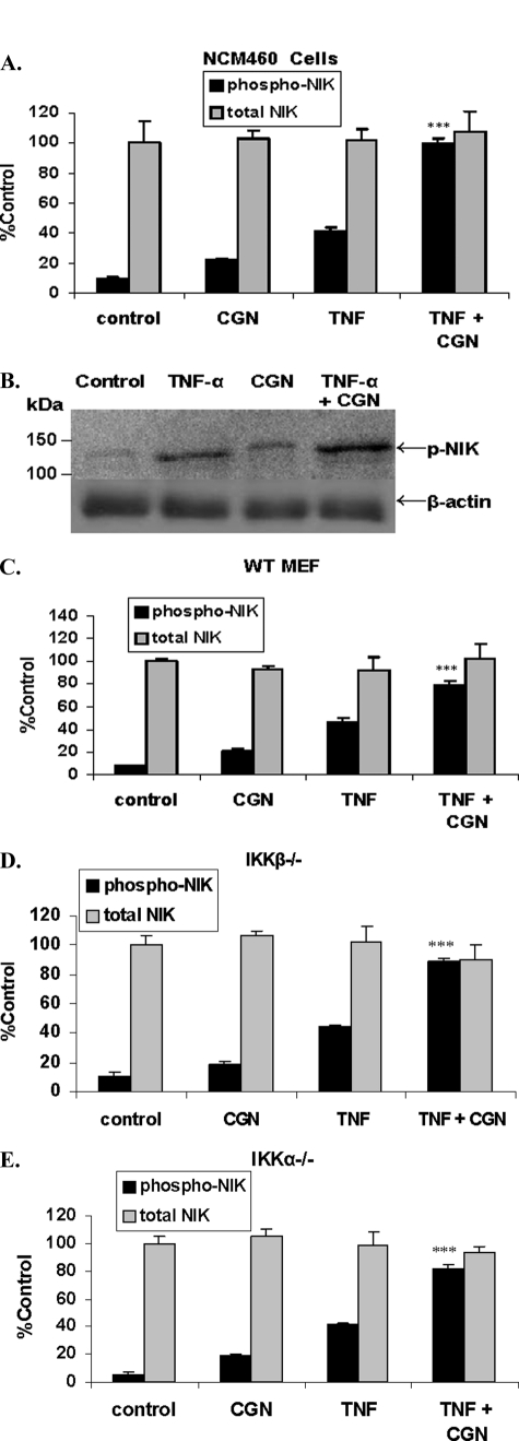
TNF-α and CGN increased NIK phosphorylation in NCM460 and MEF cells. A, combined TNF-α and CGN increased NIK phosphorylation in the NCM460 cells by ∼9.2 times the base-line value. TNF-α alone increased phospho-NIK by ∼4.3 times the base line, and CGN alone increased phospho-NIK by ∼2.2 times the base line (p < 0.001, unpaired t test, two-tailed). Total NIK content did not change. B, representative Western blot demonstrates a marked increase in phospho-NIK following exposure to TNF-α and CGN in combination. Densitometric measurements of the ratio of phospho-NIK to β-actin were averaged and indicated relative values of 1.0 for control, 4.03 ± 0.47 for TNF-α, 1.93 ± 0.14 for CGN, and 7.22 ± 0.52 for CGN and TNF-α in combination, corresponding to the increases obtained by the cell-based ELISA. C–E, both TNF-α and CGN independently induced the phosphorylation of NIK in the WT MEF (C), and the combined treatment was also synergistic in the IKKβ−/− (D) and in the IKKα−/− (E) MEF (p ≤ 0.001, unpaired t test, two-tailed). Total NIK did not change in the MEF. ***, p ≤ 0.001. Error bars, S.D.

