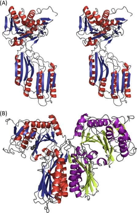
FIGURE 1.
Overall structure of V. alginolyticus PepD. A, stereo view of the subunit of V. alginolyticus PepD. Secondary structure elements are shown in red for α-helices and blue for β-strands. Gray spheres represent the zinc ions. B, ribbon diagram of the PepD dimer. The same color scheme in A is used for the left subunit, whereas the helices and strands for the right subunit are purple and green, respectively.

