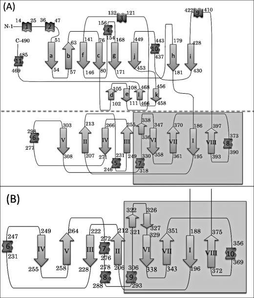
FIGURE 2.
Topological diagrams of (A) V. alginolyticus PepD and (B) the lid domain of L. delbrueckii PepV. The secondary structural elements of α-helices and β-strands are represented by ribbons and arrows, respectively. The diagram (A) has been divided into two parts, separated by the dotted line; the region with gray in the lid domain represents the extra domain.

