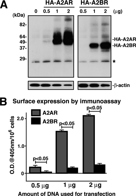
FIGURE 1.
Both the total and cell surface expression levels of A2BR are lower than those of A2AR. A, AD-293 cells were transfected with HA-A2AR or HA-A2BR with different amounts of DNA (0.5–2 μg), lysed in 1% CHAPS, and subjected to immunoprecipitation using rat mAb to HA after 72 h of transfection. The immunoprecipitates were washed and then analyzed by SDS-PAGE and immunoblotting using mouse mAb to HA. 5% of the total cell lysate was subjected to SDS-PAGE and blotted with β-actin monoclonal antibody as a loading control. Shown is a representative of three independent experiments. The asterisk indicates IgG light chain. B, AD-293 cells were transfected with HA-A2AR or HA-A2BR as in A and plated onto 96-well plates. After a 72-h transfection, cells were fixed with 4% paraformaldehyde and incubated with mouse mAb to HA followed by goat anti-mouse HRP-conjugated antibody. Cells were washed extensively with PBS, and receptors at the cell surface were determined by enzyme-linked immunoassay with ABTS solution (1 mg/ml). OD at 405 nm was measured and expressed as means (OD/106 cells) ± S.D. (n = 6).

