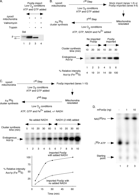
FIGURE 7.
Pos5p imported into isolated Δpos5 mitochondria stimulates Fe-S cluster biogenesis. For experiments in A–C, mitochondria were isolated from Δpos5 cells grown under reduced O2 conditions. Likewise, import and Fe-S cluster insertion assays were also performed under low O2 conditions. A, [35S]methionine-labeled Pos5p precursor protein was synthesized in reticulocyte lysate. A post-ribosomal supernatant (1 μl) containing the radiolabeled protein was incubated with Δpos5 mitochondria in the presence of ATP (4 mm) and GTP (1 mm) at 30 °C for 30 min. Valinomycin (5 μm) was included as indicated. Following import, samples were treated with trypsin (0.2 mg/ml) where indicated and analyzed by SDS-PAGE followed by autoradiography. The precursor and mature forms of Pos5p are indicated by p and m, respectively. Std indicates 10% of the precursor protein used for import experiments. B, post-ribosomal reticulocyte lysate (10 μl) with or without newly synthesized Pos5p precursor protein was added to Δpos5 mitochondria and incubated in the presence of ATP (4 mm) and GTP (1 mm) at 30 °C for 30 min. Samples were diluted with buffer A, and mitochondria with or without imported Pos5p were isolated by centrifugation. Mitochondria were resuspended in HSB buffer and supplemented with ATP (4 mm), GTP (1 mm), NADH (2 mm), ferrous ascorbate (10 μm), and [35S]cysteine (10 μCi). After incubation at 30 °C for different time periods, samples were analyzed by native PAGE followed by autoradiography. Note that [35S]methionine-labeled and imported Pos5p was not detected by native gels under these conditions. C, Pos5p was imported into Δpos5 mitochondria as in B. Mitochondria with imported Pos5p were isolated and supplemented with ATP (4 mm), GTP (1 mm), ferrous ascorbate (10 μm), and [35S]cysteine (10 μCi). NADH (2 mm) was included as indicated. Samples were incubated at 30 °C for different time periods and analyzed. The intensity of Aco1p (Fe-35S) in the presence of NADH at the 60-min time point (lane 10) was considered 100%. D, bacterially expressed and purified Pos5p was incubated with [γ-32P]ATP (0.5 μCi) plus unlabeled ATP (0.5 μm) in the presence of NADH (2 mm) at 30 °C for 5 min. Samples were analyzed by TLC followed by autoradiography.

