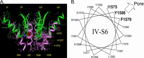
FIGURE 2.
Previously published molecular model of the voltage gated Na+ channel (2). The P loops are represented as green ribbons, S6 helices are presented as pink ribbons. A, amino acids Lys-1237 of the selectivity filter and Ile-1575 and Phe-1579 of DIV-S6 are shown by space-filling models. Amino acid numbering corresponds to the rNaV1.4 channel. Amino acid Lys-1237 of the DIII P-loop is in close spatial relationship to amino acid Ile-1575 of the DIV-S6 segment. Phe-1579 is located one helix turn internal to Ile-1575 and lines the permeation pathway in the inner vestibule. B, shown is a helical wheel representation of the DIV-S6 segment. Only amino acids downstream of Ile-1575 are shown. Amino acids in bold are considered to face the permeation pathway.

