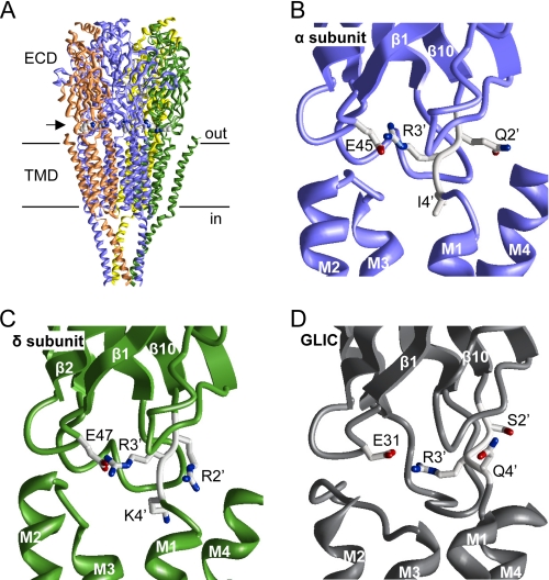
FIGURE 1.
Structure of the pre-M1 linker. A, the cryo-EM structure of the Torpedo AChR (4). The horizontal lines mark approximately the membrane. The arrow points to the location of the pre-M1 linker. The Arg3′ side chains of all subunits are space-filled. B and C, close-up view of the pre-M1 linker of the α- and δ-subunits in the Torpedo AChR. The side chains of a Glu in loop 2 and pre-M1 linker positions 2′, 3′, and 4′ are shown as sticks, where carbons, oxygens, and nitrogens are colored gray, red, and blue, respectively. The backbones of the ECD, TMD, and loops are displayed as smooth ribbons, flat ribbons, and rods, respectively. D, close-up view of the pre-M1 linker of GLIC (7). Structures were displayed using MVM (ZMM Software Inc.).

