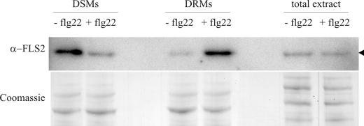
FIGURE 3.
FLS2 immunoblot analysis. Immunoblot showing reduced abundance of FLS2 in DSM and increased abundance of FLS2 in DRM fractions of flg22-treated cells. Cell cultures were treated with flg22 peptide for 10 min as described (+flg22) or remained untreated (−flg22). Subsequently, cell material was homogenized and DRMs were isolated. Total protein extracts of treated and untreated cells were used as a control to demonstrate unaltered overall FLS2 abundance, and Coomassie staining was employed to demonstrate equal loading.

