
Fig. 8.
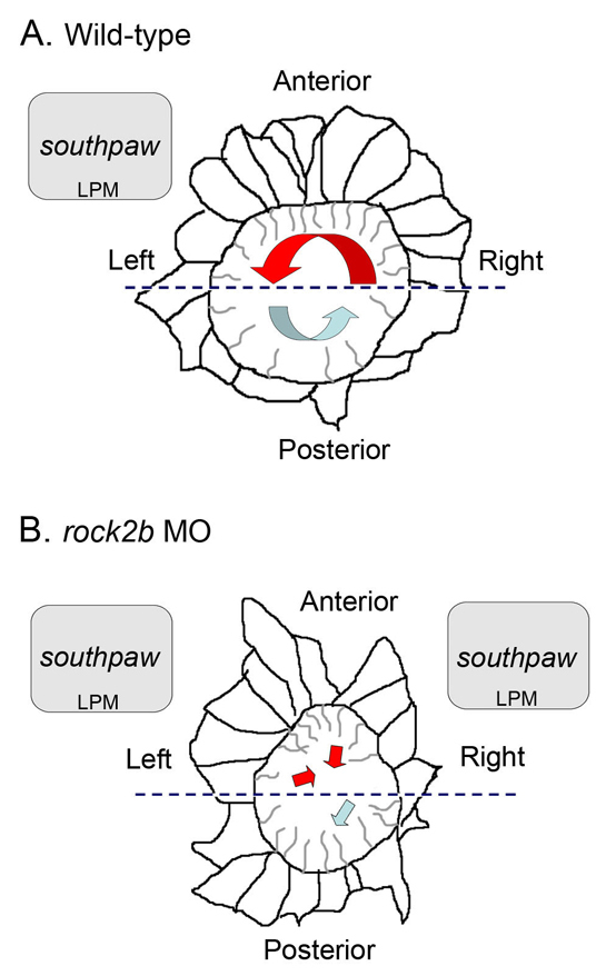
Model of the relationship between KV architecture, fluid flow and asymmetric gene expression in wild-type and rock2b knockdown embryos. (A,B) Diagrams of KV cells traced from confocal images of Tg(dusp6:memGFP)pt19 transgenic embryos with representative cilia projecting into the KV lumen. (A) In wild-type embryos, a dense packing of elongated ciliated cells in the anterior half of KV (delineated by the dashed line) drives strong leftward flow (red arrow), and fewer cilia in the posterior half of KV move fluid rightward at a slower velocity (blue arrow). We propose anterior leftward fluid flow in KV triggers asymmetric spaw expression in left LPM. (B) rock2b MO knockdown disrupted KV cell morphology and placement along the AP axis, such that ciliated cells were equally distributed in the anterior and posterior regions. This alteration of KV architecture resulted in randomized flow direction and similar flow velocities in the anterior (red arrows) and posterior (blue arrow) regions. Disruption of KV architecture and asymmetric fluid flow resulted in abnormal initiation of spaw in LPM, which was frequently bilaterally symmetric.