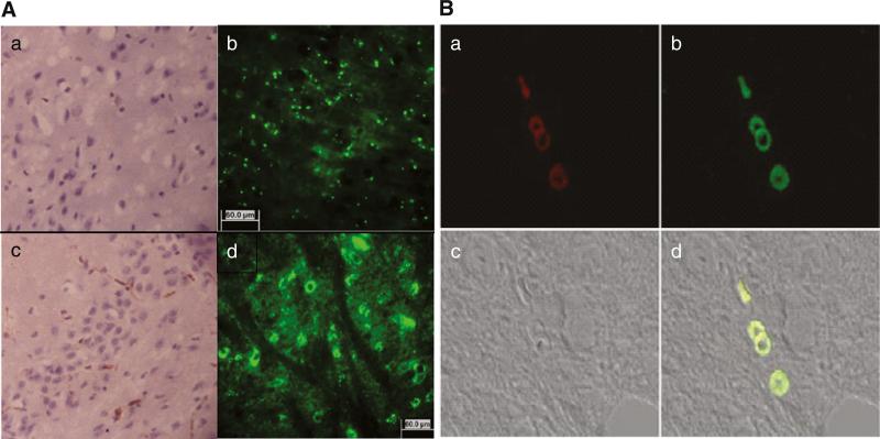
Fig. 7.
Vascular localization of GFP-expressing pDNA in mouse brain after i.v. administration. (A) anti-GFP mAb-HRP/DAB staining, and GFP fluorescence (b, d) in brain sections. a, b—non-vectorized nanoformulation, and c, d—brain-targeted ApoE-nanoformulation 48 h after i.v. injection of Balc/c mouse. (B) anti-CD34 mAb-Cy5 staining (a), GFP pDNA expression (b), bright field (c), and co-localization of the endothelial marker with GFP (d) in brain sections.

