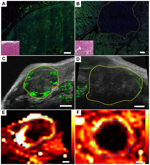
Fig. 2. Pancreatic tumors in KPC mice are poorly perfused.
Direct immunofluorescent detection of plant lectin (red) and doxorubicin (green) infused into transplanted (A) and KPC (B) tumors, along with H&E stained adjacent sections (inset). Scale bar, 200μm. Doxorubicin was effectively delivered to transplanted tumors (N=5), but poorly delivery to KPC tumors (N=4), relative to surrounding tissue. Perfusion of microbubbles (green) into transplanted (C) and KPC (D) tumors visualized by contrast ultrasonography. Transplanted tumors were well perfused (N=6) compared to KPC tumors (N=8). Tumors outlined in yellow. Scale bars, 1mm. DCE-MRI demonstrated increased perfusion and extravasation of Gd-DTPA (high delivery = white/yellow) in transplanted tumors (E, N=6) compared to KPC tumors (F, N=6). Tumors outlined in blue. Scale bars, 2mm.

