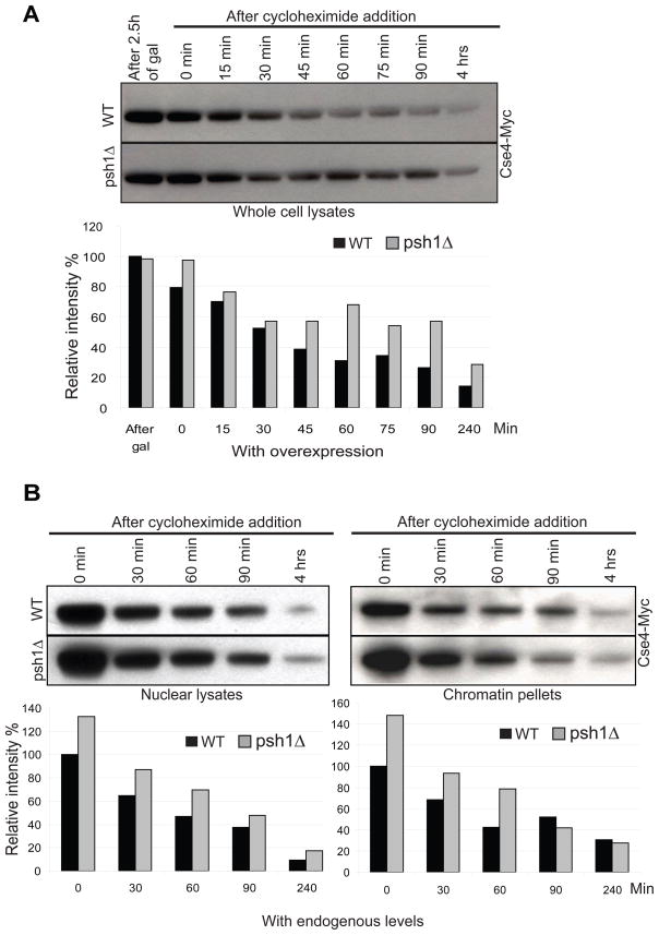
Figure 4.
Deletion of PSH1 stabilizes Cse4 protein level in vivo. Histograms indicate intensity of Cse4-Myc. A. Cse4-Myc was integrated at the URA3 locus under the control of the gal promoter. Cse4-Myc was induced by exposure to galactose for 2.5 hours in a wild-type (WT) strain or a strain in which PSH1 was deleted (psh1Δ). Cycloheximide was added and cells were collected at the timepoints indicated for Western blot analysis using an anti-Myc antibody. 70 μg of total protein was loaded per lane. B. Endogenous levels of Cse4-Myc were measured in nuclear lysates and chromatin pellets at various times following cycloheximide treatment in WT and psh1Δ strains. 30 μg of total protein was loaded per lane.

