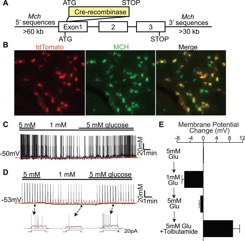
Figure 1. MCH neurons are glucose excited and express Sur1-containing KATP channels.
(A) Structure of Mch-Cre BAC transgene. (B) Double immunohistochemistry for tdTomato (red) and MCH peptide (green) in the lateral hypothalamus of Mch-Cre/lox-tdTomato mice. (C-D) Representative traces recorded in the whole-cell patch clamp mode from GFP-positive neurons of Mch-Cre/Z/EG mice. (C) Spontaneously firing MCH neuron that responded to glucose. (D) Silent MCH neuron that responded to glucose with depolarizing current injection (20pA for 3 sec with 20sec interval). (E) Effects of glucose (5mM → 1mM → 5mM) on membrane potential of “tolbutamide-responding” GFP-positive neurons of Mch-Cre/Z/EG mice. The averaged membrane potential of the last three minutes of the indicated condition from each recording was used for calculation (n=9, mean ± SEM).

