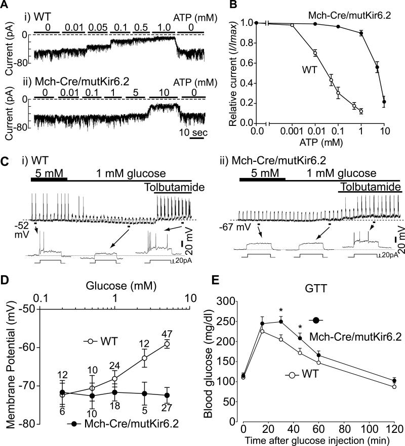
Figure 2. Expression of mutant Kir6.2 in MCH neurons blocks glucose sensing.
(A-B) Effects of ATP on KATP channel current. Recordings were performed on inside-out patches derived from GFP-positive (i.e. MCH) neurons. (A) Representative macroscopic current traces recorded at -60 mV from patches derived from i) WT (Mch-Cre/Z/EG) and ii) Mch-Cre/mutKir6.2/Z/EG mice. Concentrations of ATP are indicated above the corresponding traces. (B) Summarized effects of ATP on KATP channel current. Data was normalized to the maximal current recorded in the absence of ATP (mean ± SEM, n=5-7). (C) Effects of glucose on MCH neurons within brain slices. Representative traces recorded in the whole-cell patch clamp mode from GFP-positive neurons derived from i) WT (Mch-Cre/Z/EG) and ii)Mch-Cre/mutKir6.2/Z/EG mice. (D) Effects of glucose on membrane potential (mean ± SEM, values above each point represent the number of neurons assessed). (E) Effects of mutant Kir6.2 on glucose tolerance in intact mice. Representative glucose tolerance curves of 8-week-old male mice (mean ± SEM, n=7-10 per genotype, 2 g/Kg glucose i.p.). Asterisk, P<0.05 (unpaired t Tests), compared with wildtype littermates at a given time point.

