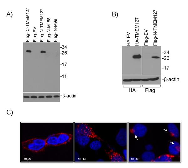
Fig.1. TMEM127 localizes to the plasma membrane and cytoplasm.
A) Western blot of HEK293 cells transfected with wild-type TMEM127 tagged with the Flag epitope at the C-terminus (C-Flag-TMEM127), N-terminus (N-Flag-TMEM127), and N-tagged TMEM127 mutants M158 (#3, Fig. 1C) and M99 (#6, Fig.1C) or empty MSCV retroviral vector (EV) and probed with a Flag antibody. A detectable product cannot be obtained from mutant constructs. β-actin was used as a loading control. B) Western blot of 293 cells transfected with TMEM127 tagged with HA at the N-terminus (HA-TMEM127) or the N-Flag-TMEM127 construct shown in (A), with respective empty vector controls, probed with an HA (left) or Flag (right) antibody. β-actin was used as a loading standard. C) Confocal microscopy of HEK293T cells expressing wild-type TMEM127 tagged with Flag on the N-terminus (left) or C-terminus (central) or with an HA N-terminal tag (right). TMEM127 immunoreactivity determined by Flag or HA (red) is present both at the plasma membrane (illustrated on the left panel) and cytoplasm, with punctate (middle and right panels) or perinuclear (right panel, arrows) signals, but is absent from the nucleus (DAPI, blue). These distinct staining patterns were observed with the various constructs in at least three independent experiments (Suppl. Fig. 4B). Plasma membrane-associated TMEM127 distribution was detected on average in 38% (±13%, n=200) of cells under regular culture conditions. A similar variance in the percentage of plasma membrane associated-signal was noted for each of the constructs.

