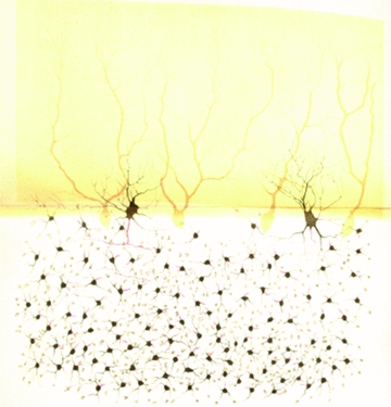
Figure 3.

The relationship between Golgi cells and granule cells Granule cells largely exceed in number the Golgi cells and are much smaller (Purkinje cells are distinctly shown in the background). Therefore a Golgi cell can innervate several granule cells lying within their axonal plexus, capturing another fundamental feature of the granular layer organisation. (Fragment of a rabbit cerebellar convolution (vertical section); Table XIX, Opera Omnia, Golgi, 1903). ‘This drawing was specifically made to illustrate the granular layer. … The so-called granule cells look like nervous cells with a globose shape, really small and equipped with 3, 4, 5 or even 6 prolongations, among which just one has the features of nervous prolongation (the nervous prolongation is just outlined, red thread). Prolongations, which it seems to be correct to name protoplasmatic, even if they appear slightly different from other gangliar cells’ prolongations, end up with a small granulous mass, towards which neighbouring granule cells’ prolongation often converge. In the region in which the granular layer merges into the molecular layer, two large cells are drawn. These are placed laterally and differ from Purkinje cells for the cell body shape, for the way of branching of their protoplasmatic prolongations and, overall, for the very different organisation of the nervous prolongation. … These two large cells are of the same type of the ones already illustrated in Tables XIV and XVII.’ Translated from Opera Omnia (Golgi, 1903).
