
Figure 3. Roles of phospholipase C and intracellular Ca2+ stores in ADP-induced currents.
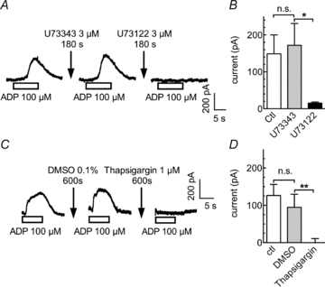
A and B, after an initial application of ADP, cells were treated for 420 s with control solution followed by a 180 s application of U73343 (3 μm) and a second ADP application. This was followed by a 420 s washout, a 180 s treatment with U73122 (3 μm), and a third ADP application. A, sample currents recorded from a single PC12 cell. B, peak current amplitudes in the absence or presence of either U73343 or U73122 (n= 4); *significant difference at P < 0.05. C and D, cells were continuously superfused with control solution (ctl), DMSO (0.1%) or 1 μm thapsigargin, each for 10 min. Outward currents were elicited by 10 s applications of ADP at the end of these treatments. Original current traces (A) and peak amplitudes of the currents (B) evoked by ADP under control conditions (ctl) and in the presence of either 0.1% DMSO or 1 μm thapsigargin (**P < 0.01; n.s. no significant difference; n= 5).