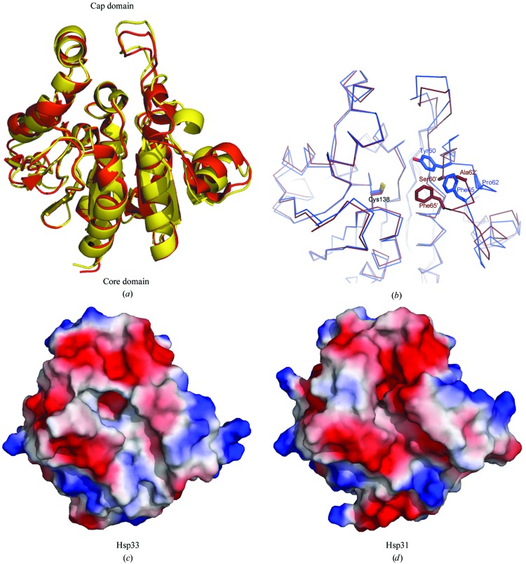
Figure 4.
Structural comparison of yeast Hsp33 and Hsp31. (a) Superimposition of the structures of Hsp33 (yellow) and Hsp31 (red). (b) Comparison of the active-site pocket between Hsp33 (blue) and Hsp31 (brown). The different residues are presented as sticks. The electrostatic surface of (c) Hsp33 and (d) Hsp31, showing the differences in the size and orientation of the active pocket. All figures were made with PyMOL (DeLano, 2002 ▶).

