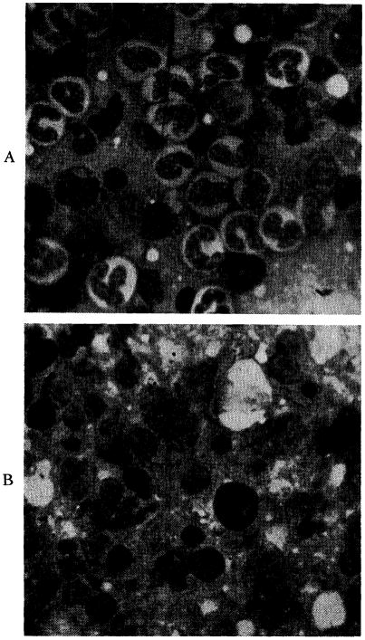
Fig. 13.
A, bone marrow from normal dog showing active granulopoiesis and erythropoiesis (magnification × 900). B, marrow from Dog No. 26, showing a cellular specimen with extensive replacement of normal myeloid elements by a relative and absolute increase in lymphocytes, reticulum cells and plasma cells (magnification × 900).

