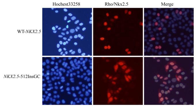
Fig. 3.
Cellular localization of wild type NKX2.5 (WT) and mutant NKX2.5 generated by mutation c.512insGC. HeLa cells were transfected with pWT-NKX2.5 and pNKX2.5-512insGC and stained with a goat anti-NKX2.5 antibody as the primary antibody and rhodamine-labeled donkey anti-goat antibody as the secondary antibody (red signal). Nuclei were stained with Hochest 33258 (blue signal). Wild type NKX2.5 was localized completely into the nucleus, whereas the mutant NKX2.5 was localized in both nucleus and cytoplasm.

