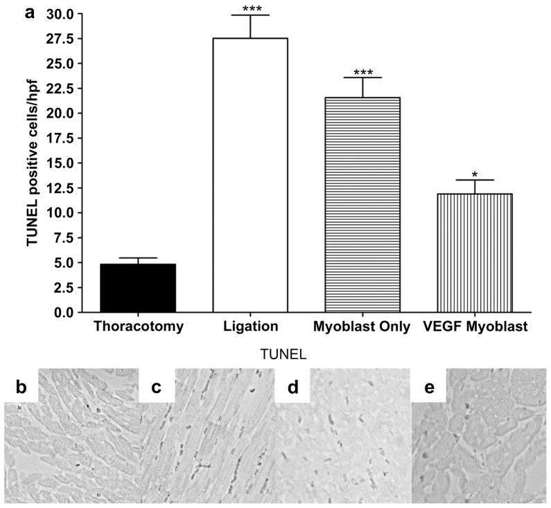
Figure 6.
TUNEL staining for apoptotic activity, 4weeks after surgery. (a) TUNEL staining for apoptotic cells 4weeks after surgery. TUNEL-positive apoptotic cells were counted in the five random high-power fields (hpf) per animal in the ischemic border zone. The average number of apoptotic cells was statistically greater between thoracotomy control and all groups with less significance between thoracotomy and the VEGF myoblast group. (P<0.001 thoracotomy vs ligation and myoblast only; P<0.05 between thoracotomy and VEGF myoblast group.) The VEGF myoblast group had significantly fewer apoptotic cells than the ligation group (P<0.001) and the myoblast only group (P<0.01). There was no significant difference between the myoblast only treatment group and ligation controls. Representative micrographs of apoptotic nuclei by TUNEL staining in (b) thoracotomy control group, (c) ligation, (d) myoblast only injection group and (e) VEGF165-transfected myoblast injection group. All data are presented as mean ± SEM (n=5 thoracotomy, n=7 ligation, n=7 myoblast only, and n=7 VEGF myoblast group).

