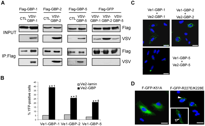
Figure 4. The in vivo homodimerization of GBPs differentially influences their intracellular localization.
(A) GBP-1, GBP-2 and GBP-5 are able to homodimerize in vivo. HeLa cells were co-transfected with Flag-GBPs or Flag-GFP together with VSV-GBPs or empty control vector (CTL) as indicated. Protein extracts were immunoprecipitated with an anti-Flag affinity gel and subjected to western blot analysis. For each co-transfection, cell lysates (10 µg, INPUT) and IP eluates (1∶4 for Flag-detection and 3∶4 for VSV detection) were analyzed. (B) Quantitative evaluation of the bi-molecular fluorescence complementation assay. Venus1 plasmids encoding various GBPs were co-transfected with matching Venus2-GBP (black bars) or Venus2-lamin (gray bars) encoding plasmid into HeLa cells. The number of fluorescent cells was determined by FACS analysis after 72 h. (C) Bi-molecular fluorescence complementation assay. HeLa cells were transfected with the plasmids expressing GBP-1, -2 or -5 fused with Venus1 or VSV-Venus2, respectively. Nuclei were counterstained with DAPI. (D) HeLa cells were transfected with different mutants of GBP-1 fused to GFP. Flag-GFP-GBP-1-K51A is defective in nucleotide-dependent oligomerization. Flag-GFP-GBP-1-R227E/K228E is constitutively dimeric and localizes in vesicle-likes structures (solid arrowhead) or at the plasma membrane (open arrowhead). Nuclei were counterstained with DAPI. Scale bars = 25 µm.

