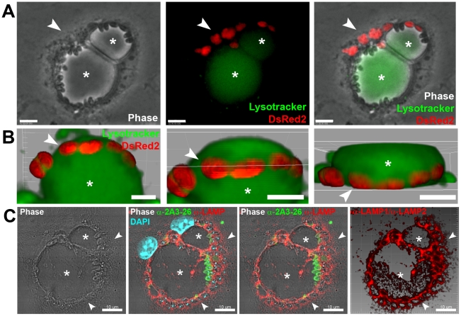
Figure 1. L. major amastigote PVs did not fuse with L. amazonensis vacuoles.
(A) Macrophages were previously infected for 48 h with L. amazonensis-WT and then superinfected with L. major-DsRed2 amastigotes for additional 72 hours. Image shows a macrophage loaded with Lysotracker (green) and hosting the two parasite species under phase contrast channel (Ph2), fluorescence channels (Lysotracker and DsRed2), and merged channels, respectively. Asterisk indicates L. amazonensis PV and arrowheads indicate L. major-DsRed2 amastigotes (red). Bars = 10 µm. (B) Live multidimensional imaging of coinfected macrophages. Asterisk indicates L. amazonensis-WT PV stained with Lysotracker (green), surrounded by membrane-bound PVs with weak Lysotracker signal which shelter L. major-DsRed2 amastigotes (red). Multidimensional images were constructed by Imaris blend filter and each image represents a rotation of approximately 45°. Bars = 5 µm. (C) Immunolocalization of LAMP1/LAMP2 proteins in superinfected macrophages. Leishmania major amastigotes were sheltered by tight LAMP1/LAMP2-positive PVs (arrowheads), close to large recipient L. amazonensis PVs, indicated by asterisks. Image was acquired 11 days after L. major-DsRed2 amastigote addition. LAMP immunolabeling in red, 2A3-26 antibody (specific for L. amazonensis amastigotes) immunolabeling in green, DAPI staining in blue. Images are disposed as phase contrast (Ph3), phase contrast with RGB fluorescence channels, phase contrast with RG channels and 3D reconstruction of red channel with Imaris blend filter. Bars = 10 µm.

