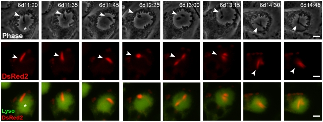
Figure 4. L. major promastigotes multiply inside chimeric PVs.
Time-lapse recording of macrophages infected with L. amazonensis-WT for 48 hours and superinfected with metacyclic-enriched L. major-DsRed2 promastigotes. Image acquisition started 6 days after L. major-DsRed2 promastigote addition. Division of L. major-DsRed2 promastigote (arrowheads) inside L. amazonensis-WT PV (asterisk) was documented. The figure shows phase contrast (Ph2) in the first row, DsRed2 signal in the second, and Lysotracker merged with DsRed2 signal in the third. Time after promastigote addition is shown (d:h:min). Scale at 10 µm.

