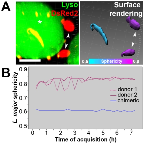
Figure 5. Multidimensional image of metacyclic-enriched L. major-DsRed2 promastigotes in superinfected macrophages.
(A) On the left, multidimensional image of a chimeric PV (asterisk) and L. major-DsRed2 parasites sheltered by unfused donor PVs (arrowheads); Imaris MIP filter. On the right, surface rendering of parasites through DsRed2 channel allowed the software to assign a colorimetric scale to each L. major-DsRed2 parasite: it displays the sphericity parameter, ranging from cyan (less spherical, 0.5) to magenta (more spherical, 0.8); Imaris blend filter. Images were acquired 24 hours after addition of L. major-DsRed2 metacyclic-enriched promastigotes to macrophages. Bar = 10 µm. (B) Sphericity measurements during coinfection, presented by L. major-DsRed2 parasites hosted within unfused donor PVs (magenta lines) or within chimeric PV (blue line). Acquisition of multidimensional images started 12 hours after L. major-DsRed2 metacyclic-enriched promastigotes were added to macrophages.

