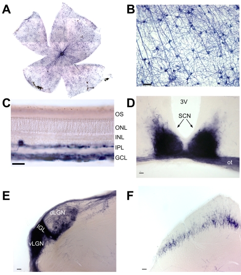
Figure 1. Genetic labeling of melanopsin RGCs and their projections.
(A) Alkaline phosphatase (AP) stained mRGCs in Opn4Cre;Z/AP mice are uniformly distributed across the retina (1,556±72; mean ± SD, n = 4). (B) AP labels the soma, dendrites, and axons of mRGCs. (C) Labeled cell bodies are restricted to the ganglion cell layer (GCL) and inner nuclear layer (INL), with dendrites in both sublaminae of the inner plexiform layer (IPL). (D–F) AP-stained coronal brain sections demonstrate dense mRGC innervation of the suprachiasmatic nuclei (SCN, D), intergeniculate leaflet (IGL, E), ventral and dorsal lateral geniculate nuclei (v/dLGN, E), and sparse innervation of the superior colliculus (F). 3V, third ventricle; ot, optic tract. Scale bars represent 50 µm (B, D–F) and 25 µm (C).

