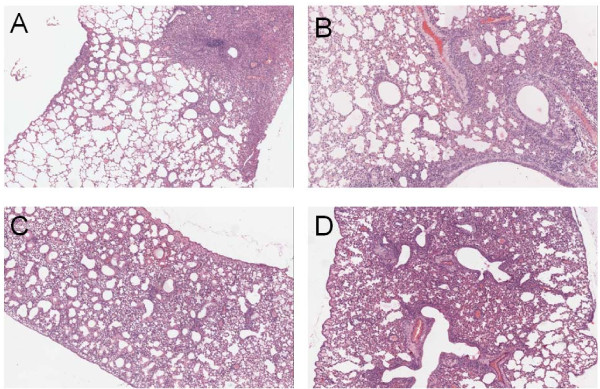
Figure 3.
Pathological analysis of the lung tissues of challenged BALB/c mice. The images were obtained on an OLYMPUS BX-50 light microscope with 40× magnification. (A) Lung tissues of mice challenged with A/California/04/2009 virus; (B) Lung tissues of mice challenged with A/California/07/2009 virus; (C) Lung tissues of mice challenged with mut-A/California/04/2009 virus; (D) Lung tissues of mice challenged with mut-A/California/07/2009 virus.

