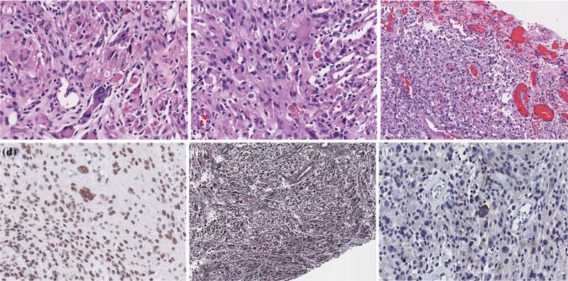
Fig. 2.
Photomicrographs of the right temporal lobe pleomorphic xanthoastrocytoma, showing compact regions with cellular pleomorphism and scattered eosinophilic granular bodies (a,b), a leptomeningeal compact region with perivascular and interstitial lymphocytes, xanthoma cells and pleomorphic cells (c), retained SMARCB1/BAF47 (INI-1) nuclear staining (d), reticulin staining of the superficial component with reticulin fibers around individual cells and clusters of cells (e) and synaptophysin staining showing positivity in some of the large pleomorphic cells (f). Original magnification, 200×

