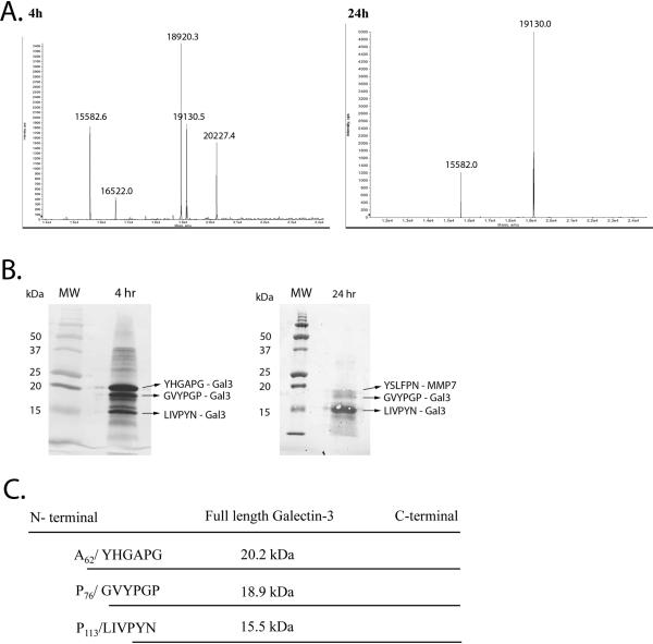
Figure 2. Identification of MMP7 induced galectin-3 cleavage site.
A. Mass spectra of recombinant galectin-3 and MMP7 reaction products. Recombinant MMP7 and galectin-3 were incubated together for 4 and 24 hours and mass spectrometry performed. B. Coomassie Brilliant Blue staining of the 4 and 24 hr reaction mixtures utilized for subsequent N-terminal sequencing. C. Schematic representation of MMP7-directed galectin-3 cleavage sites for the 20.2, 18.9 and 15.5 kDa galectin-3 fragments. (figure not to scale.)

