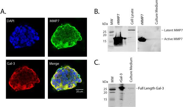
Figure 4. Galectin-3 and MMP7 are expressed in T84 cells.
A. T84 cell colonies were stained for anti-galectin-3 C-terminus (red), MMP7 (green) and DAPI (blue) and visualized by confocal microscopy. Predominant MMP7 and galectin-3 staining were seen in the periphery with co-localization (yellow) seen in the merged image. B. Western blot analysis detected the 28 kDa pro-MMP7 in T84 cell lysates and culture medium supernatants. C. Western blot analysis detected the full length galectin-3 protein in the T84 culture medium supernatants.

