
Figure 2.
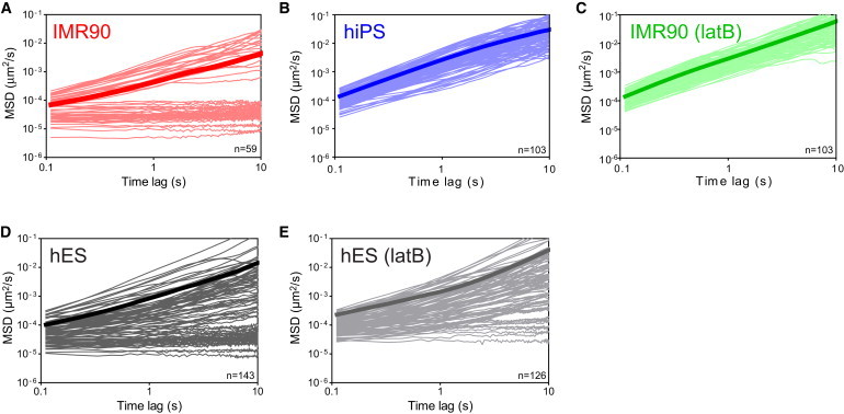
MSDs of beads in parental human fibroblasts, derived hiPS cells, and hES cells. (A–E) MSDs of individual beads embedded in the cytoplasm of parental IMR90 fibroblasts (A, n = 59), derived hiPS cells (B, n = 103), latB-treated IMR90 fibroblasts (C, n = 103), hES cells (D, n = 143), and latB-treated hES cells (E, n = 126). Time-lag-dependent MSDs exhibiting a slope <1 on a log-log plot (A, D, and E) are subdiffusive and indicative of a cytoplasm that is locally elastic, whereas slopes of 1 indicate a diffusive environment. Note the complete absence of subdiffusive MSDs in hiPS cells and the reduction of subdiffusive MSDs in latB-treated hES cells relative to untreated hES cells.