
Figure 4.
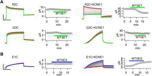
KCNE1 association enhances extracellular MTS accessibility to the VSD. (A) MTSET+ modification of R2C and Q3C in the absence and presence of KCNE1. Oocytes were held at −80 mV for 32 s and repeatedly pulsed to +40 mV for 5 s, and repolarized at −40 mV for 3 s. MTSET+ reduces R2C current only in the presence of KCNE1. Q3C current is unchanged by MTSET+. Scale: R2C 4 μA, R2C+KCNE1 30 μA, Q3C 1.5, Q3C+KCNE1 15 μA; 2 s. The peak current amplitude after each voltage pulse is plotted against time. (B) MTSES− modification of E1C in the absence and presence of KCNE1 using the voltage protocol in A. MTSES− enhances E1C current only in the presence of KCNE1. Scale: E1C 2 μA, E1C+KCNE1 4 μA; 2 s.