
Figure 2.
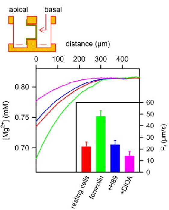
Inhibition of KCC1 has a minor effect on osmotic water flux through IMCD cells in the absence of apical AQP2. A scheme of the experimental setup is shown at the top. The confluent cell monolayer on a Transwell filter is mounted vertically in a Teflon chamber. The Mg2+-sensitive microelectrode and a reference electrode are placed in the hyperosmotic (basolateral) compartment. The osmotic pressure was induced by 450 mM PEG600. The resulting water flux led to Mg2+ dilution, which allowed calculation of the apparent water permeability Pf in the steady state (inset). Therefore, we took both the near-membrane dilution of PEG600 and the decrease of the PEG and Mg2+ diffusion coefficients (compare Fig. S1) into account. First we measured the Mg2+ dilution in the immediate membrane vicinity of resting cells. To show that the cells are functionally active, we then added forskolin, which induced exocytic insertion of aquaporin-2 (AQP2) water channels into the apical membrane. As a result Pf, and thus, the Mg2+ dilution, increased. Subsequent application of the exocytosis inhibitor H89 (50 μM) resulted in endocytic retrieval of AQP2, and thus, reestablished Pf of resting cells. The subsequent addition of DIOA (15 μM) induced a very modest decrease of Pf due to KCC1 inhibition. (Inset) Average Pf values for six runs of the above-described sequence of additions. The water fluxes values for the representative records shown were 0.9, 1.9, 0.9, and 0.6 μmol cm2 s−1 for, respectively, the control monolayer and the monolayers subsequently treated by forskolin, H89, and DIOA.