
Figure 5.
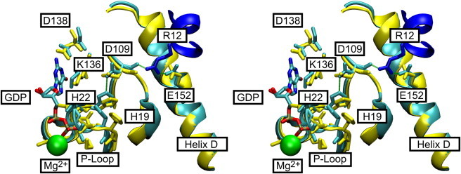
Structure of the nucleotide-binding pocket (stereo view). (A) H-bonding interactions between Asp109 and His22: detailed view of the nucleotide interactions in the EF-Tu•GDP (yellow) and EF-Tu•EF-Ts (cyan for EF-Tu and blue for EF-Ts) complexes. Residues involved in nucleotide binding are highlighted. Arg12 of EF-Ts is depicted in dark blue, and Mg2+ is represented by a green sphere. H-bonds of the proposed H-bond relay are indicated in green. The figure was prepared with VMD (17) using coordinate sets 1EFC and 1EFU in the PDB (3,24).