
Figure 2.
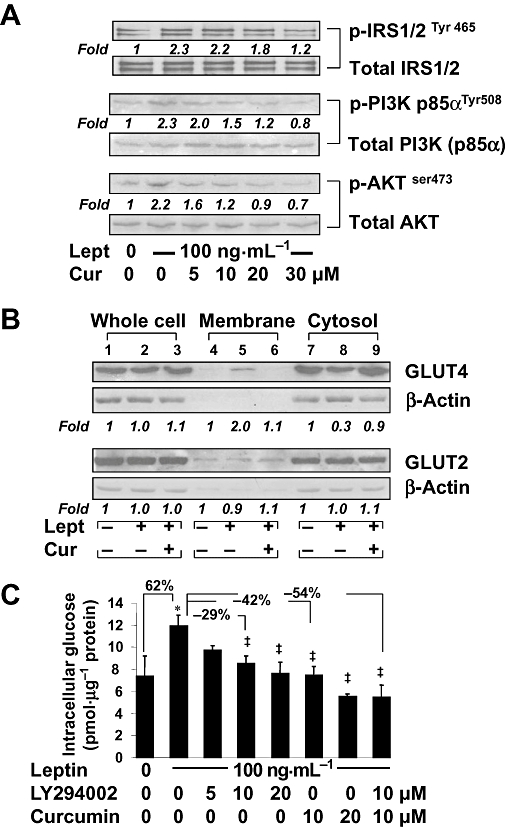
Curcumin interrupted the leptin-activated IRS/PI3K/AKT signalling pathway in cultured HSCs, leading to inhibition of the GLUT4 membrane translocation and to reduction of intracellular glucose. Serum-starved HSCs were pretreated with or without curcumin at indicated concentrations for 1 h prior to the stimulation with or without leptin (100 ng·mL−1) in serum-free media for additional 30 min. Western blotting analyses were conducted (A,B). Representative blots are shown from three independent experiments. Italic numbers beneath blots are fold changes in the densities of the bands compared to the control without treatment in the blot (n = 3), after normalization with the internal invariable control. Because of the limited space, standard deviations are not presented. (A) Detection of phosphorylated IRS1/2, PI3K and AKT. The total protein for each corresponding phospho-protein was used as an internal control for equal loading. (B) Evaluation of the abundance of GLUT2 and GLUT4, respectively, in the fractions of whole cells, membrane and cytoplasm of HSCs. (C) Serum-starved HSCs were pretreated with or without the PI3K/AKT inhibitor LY294002 at indicated concentrations, or with curcumin (20 µM), for 1 h prior to the stimulation with or without leptin (100 ng·mL−1) in serum-free media for additional 30 min. Levels of intracellular glucose were determined. Values were expressed as pmol glucose·µg−1 protein and presented as means ± SD. *P < 0.05 versus the untreated control; ‡P < 0.05 versus cells treated with leptin alone.