
Figure 3.
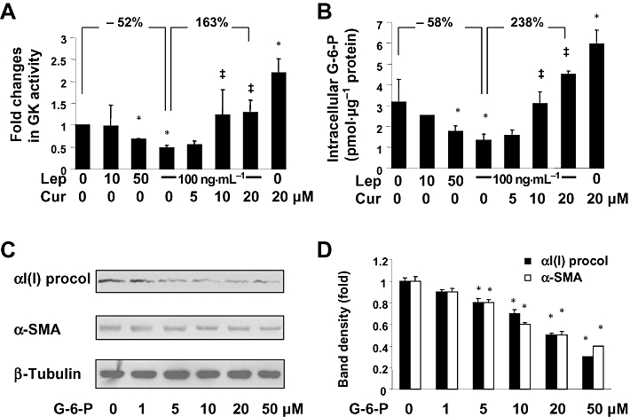
Curcumin eliminated the inhibitory effect of leptin on the activity of GK and increased intracellular G-6-P in activated HSCs in vitro. (A,B) Serum-starved HSCs were pretreated with curcumin at indicated concentrations for 1 h prior to the stimulation with or without leptin (100 ng·mL−1) in serum-free media for additional 30 min. (A) Analyses of GK activities. Values were expressed as fold changes (means ± SD), compared with the untreated control (n = 3). *P < 0.05 versus the untreated control; ‡P < 0.05 versus cells treated with leptin at 100 ng·mL−1 alone. (B) Analyses of levels of intracellular G-6-P. Values were expressed as pmol G-6-P·µg−1 protein (means ± SD) (n = 3). *P < 0.05 versus the untreated control; ‡P < 0.05 versus cells treated with leptin at 100 ng·mL−1 alone. (C) Western blotting analyses of αI(I)procollagen (procol) and α-SMA in HSCs treated with G-6-P at different concentrations as indicated in serum-depleted media for 24 h. β-Tubulin was used as an invariant control for equal loading. Representatives were presented from three independent experiments. (D) The Western blotting analyses were summarized after normalization with β-tubulin. Variations in the band density were expressed as fold changes compared to the untreated control in the blot (means ± SD., n = 3). *P < 0.05 versus the untreated control.