
Figure 5.
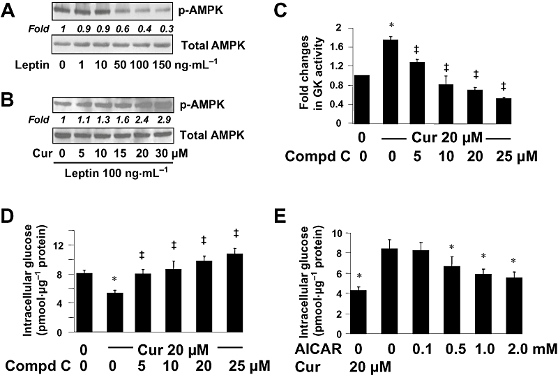
Curcumin activated AMPK activity, which resulted in the increase in the activity of GK and in the reduction in the level of intracellular glucose in cultured HSCs. (A,B) Serum-starved HSCs were pretreated with or without curcumin (0–30 µM) for 1 h prior to the stimulation with leptin at indicated concentrations in serum-free media for additional 30 min. Levels of p-AMPK were evaluated by Western blotting analyses. Total AMPK was used as an invariant control for equal loading. Representative blots are presented from three independent experiments. Italic numbers beneath blots were fold changes in the densities of the bands compared to the control without treatment in the blot (n = 3), after normalization with total AMPK. (A) Cells were stimulated with leptin only. (B) Cells were pretreated with curcumin prior to the stimulation with leptin (100 ng·mL−1). (C,D) Serum-starved HSCs were pretreated with or without curcumin at 20 µM for 1 h prior to the exposure to the AMPK inhibitor compd C at various concentrations in serum-depleted media for additional 30 min. (C) Analyses of GK activities. Values were expressed as fold changes compared with the untreated control (means ± SD) (n = 3). *P < 0.05 versus the untreated control; ‡P < 0.05 versus cells treated with curcumin alone. (D) Analyses of levels of intracellular glucose. Values were expressed as pmol glucose·µg−1 protein (means ± SD) (n = 3). *P < 0.05 versus the untreated control; ‡P < 0.05 versus cells treated with curcumin alone. (E) HSCs were treated with the AMPK activator AICAR at various concentrations, or with curcumin at 20 µM, for 1.5 h. Levels of intracellular glucose were determined. Values were expressed as pmol glucose·µg−1 protein (means ± SD) (n = 3). *P < 0.05 versus the untreated control (the second column).