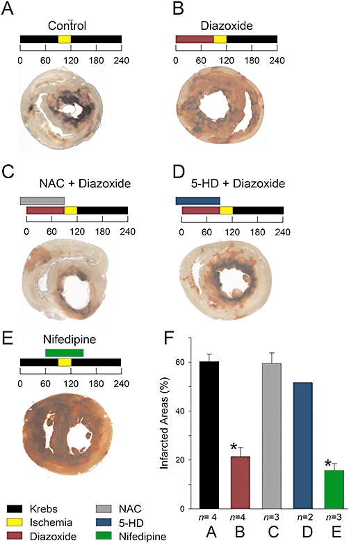
Figure 9.

PPC and infarction areas. A–E, infarction areas (light areas) after severe ischaemia in cross-sections of isolated ventricles under conditions as indicated above. Numbers are time in minutes. Concentrations were 100 µM diazoxide, 4 mM NAC, 100 µM 5-HD, and 30 nM nifedipine. Ventricles were dyed with 2,3,5-triphenyltetrazolium chloride. F, infarction areas under indicated conditions corresponding to panels A–E. Data are the mean relative infarcted area (±s.e.) with the number of experiments indicated below. Infarcted area values from hearts perfused with diazoxide and nifedipine were significantly lower than those from control hearts (*P < 0.05).
