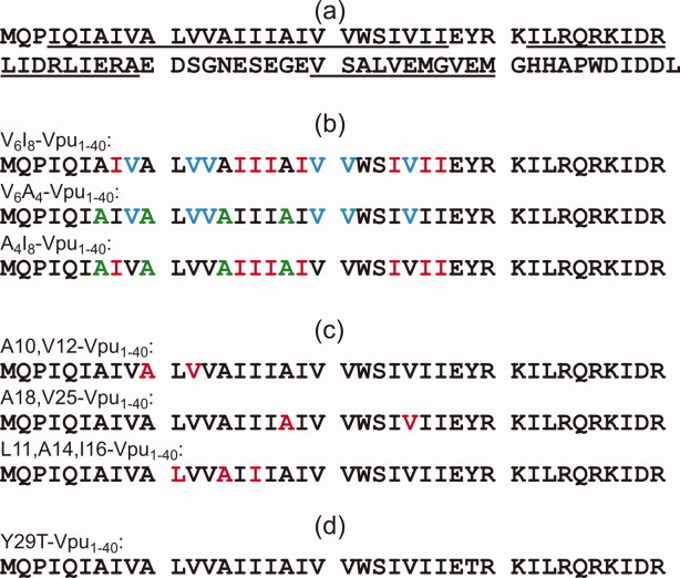
Figure 1.

(a) Full-length HIV-1 Vpu sequence, with three helical segments underlined. Location of N-terminal, transmembrane helical segment is based on solid-state NMR data of Sharpe et al.17 Central and C-terminal helical segments are based on data of Opella and coworkers.1,11 (b) Vpu1–40 peptides used in 2D CHHC measurements of intermolecular contacts, shown in Figure 8. Samples contain 1:1 mixtures of peptides with uniform 15N,13C-labeling of six Val (cyan), eight Ile (red), or four Ala (green) residues. (c) Vpu1–40 peptides used in DIPSHIFT measurements of molecular orientation, shown in Figures 9 and 10. Uniformly labeled residues are indicated in red. (d) Y29T-Vpu1–40 peptide used in analytical ultracentrifugation and PICUP experiments, shown in Figures 2–7. [Color figure can be viewed in the online issue, which is available at wileyonlinelibrary.com.]
