
Figure 3.
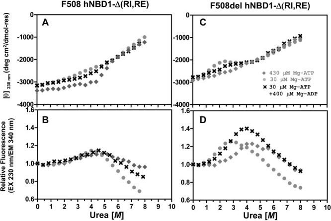
Effects of Mg-ADP versus Mg-ATP on isothermal urea denaturation of hNBD1-Δ(RI,RE). Denaturation of the F508 (left) or F508del (right) domain was conducted at 25°C under conditions identical to Fig. 1 and monitored using CD (top) and trp fluorescence (bottom, with 230 nm excitation and 340 nm emission). Proteins were diluted into Standard Stabilizing Buffer containing 400 μM Mg-ADP (crosses), 400 μM Mg-ATP (gray diamonds), or no additional nucleotide (gray closed circles). Because purified proteins are stored in Standard Stabilizing Buffer containing Mg-ATP, they introduce 30 μM Mg-ATP into every experimental sample in addition to any nucleotide present in the measurement buffer. The final Mg-ATP concentration in each experiment is indicated in the legends.