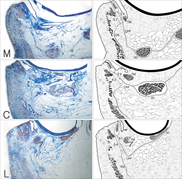
Fig. 6.
Three parasagittal sections on the medial limbus (M), the midpupillary line (C) and the lateral limbus (L). 10 µm section, Masson-trichrome stain, ×10 magnification. The head of the capsulopalpebral fascia (CPF) splits open superiorly and inferiorly and then it wraps around the inferior oblique muscle (IO) and meets anteriorly. The CPF inserted in to the inferior border of the tarsus, and then it merges with the anterior border of the inferior tarsal muscles (TM). The orbital septum (OS) blended with CPF most closely at 3.7~5.4 mm beneath the lower tarsal border: and differently at 3.7±0.7 mm on the medial limbus line, 4.3±0.8 mm on the midpupillary line and 5.4±1.0 mm on the lateral limbus line. A distinct bundle of capsulopalpebral fiber was seen about 3 mm below the lower border of the tarsal plate, 2.9±0.6 mm on the medial limbus line, 2.9±0.6 mm on the midpupillary line and 3.1±0.9 mm on the lateral limbus line (Hwang et al., 2006).

