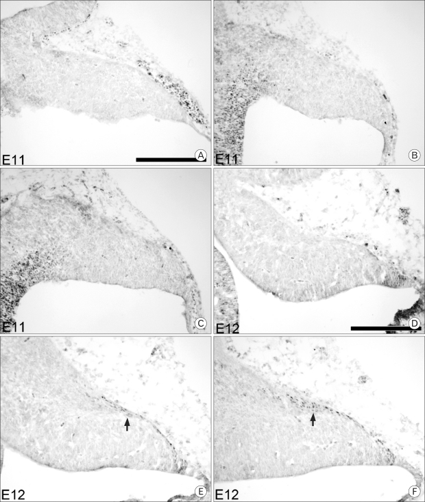
Fig. 1.
Pax6 expression at embryonic day E11 and E12 in the cerebellum. The top row shows medial sections, the middle row shows paramedial sections and the bottom row shows lateral sections that correspond to mediolateral locations. Immunocytochemistry with an antibody against Pax6 was performed on sagittal sections. No Pax6 expression is observed at E11 (A~C). At E12 (D~F), cells immunoreactive for Pax6 are first seen in a single layer on the dorsal surface, as well as a small group of cells at the rostral end of the layer (arrow). Scale bar is 250 µm.

