Fig. 3.
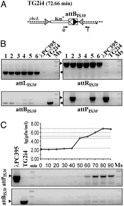
Integration and excision of λPC395 in the chromosomal attBIS30 site. (A) Location of the integrated attBIS30 in the E. coli genome. The shaded arrowheads bracketing the KmR gene with the IS30 IR-IR junction represent the ends of Tn10 minitransposon (see Materials and Methods). Thin arrow, yhcL gene; e and f, primers used in PCRs. (B) PCR analysis of six integrant lysogens. Lanes 1-6 show the amplification products obtained with the primer combinations of e-f for attBIS30 (base pairs 571), c-d for attPIS30 (base pairs 890; see Fig. 2B), e-d for attLIS30 (base pairs 512), and c-f for attRIS30 (base pairs 943). The weak attBIS30-specific signals suggest that these clones showed spontaneous excision similarly to a wild-type λ lysogen. Positive signals for attPIS30 in lanes 2 and 5 may also be derived from imperfect phage repression. (C) Excision of λPC395 during lytic phase of a lysogen. The excision was assayed by titration of phages and PCR detection of attBIS30 and attPIS30.

